![]()
Patients affected by canine pododermatitis are presented commonly in clinical small animal practice. The condition may be present on a single claw, a single paw, or multiple paws. While the condition may affect the paw(s) only, it may be accompanied by additional cutaneous symptoms at other sites, depending on the underlying cause. Pododermatitis is defined as inflammation of the skin of the paw. This definition includes all potentially affected pedal tissues such as interdigital spaces, footpads, nail folds, and nails. It is important to remember that pododermatitis is a clinical presentation, not a final diagnosis.
Nature of the clinical signs exhibited and chronicity of the problem is dependent on multiple factors including the primary underlying cause and presence of secondary infection. It would not be wrong to liken pododermatitis to otitis, with presence of multiple factors affecting each both conditions.
With otitis externa, we are always mindful of the presence of primary factors responsible for the condition, as well as perpetuating factors and predisposing factors that may make otitis more likely to develop and difficult to resolve. Canine pododermatitis is not dissimilar, with presence of multiple factors that may make resolution complicated.
Multiple factors are usually at play such as paw conformation (that can be breed dependent), primary underlying conditions (such as allergies), as well as secondary perpetuating infection and chronic changes to local cutaneous health. As in otitis patients, clinical findings related exclusively to the paws can appear to be the same despite the various underlying conditions and secondary problems associated with pododermatitis.
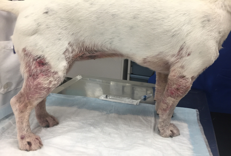
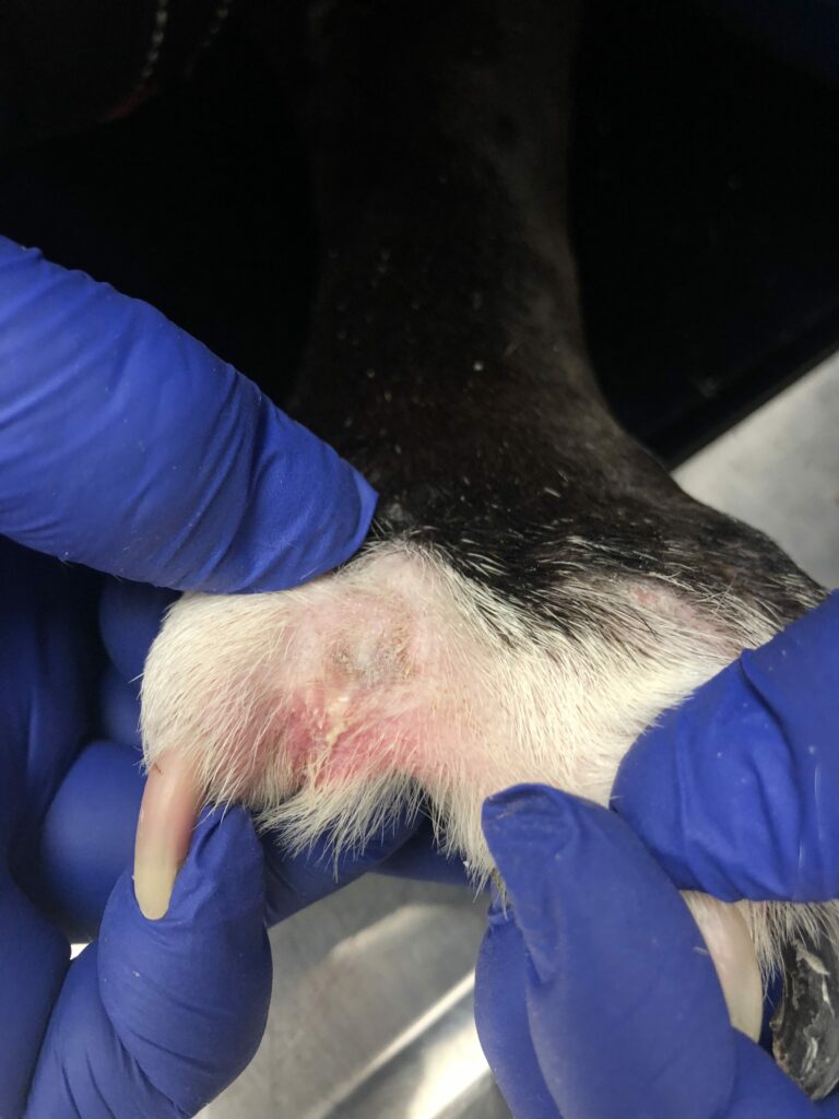
Affected paws may be affected with pruritus, erythema, edema with or without nodules, paronychia, alopecia, ulceration, paw pad involvement, comedones, swelling, and draining exudates. The skin may be moist from constant licking and/or infection, and varying degrees of pain, pruritus, and lameness may be evident.
Some Common Pedal Conditions in Dogs that Veterinarians may Diagnose
- Pedal erythema – Interdigital erythema of pedal skin is probably the most common clinical presentation of canine pododermatitis. While most patients are not presented for erythema itself; when presented for pruritus or an allergic history, erythema is often noted on one or more paws. Erythema is indicative of an active inflammatory process due to allergies, including epicutaneous absorption of allergens (atopic dermatitis), food allergy, and contact allergy. Secondary infection can also lead to erythema of pedal skin, with or without presence of primary allergies.
- Pedal pruritus – Another very common presentation of pedal disease is itchiness. It is well documented that itchiness of paws presents as excessive licking or chewing of paws. Pedal pruritus is commonly seen with allergies in pets, as well as related to infection of the paws due to Malassezia yeast or bacterial pododermatitis. Often, combination of an underlying allergy and a secondary infection may be present in a pet presented with pedal pruritus.
Often, pet families may mistakenly associate paw licking or biting with anxiety! This is likely due to the incorrect correlation with nail-biting in humans with stress or anxiety. Pets itch their paws most often due to allergies and presence of paw infection.
- Broken nails – Primary nail trauma is commonly seen in active dogs that may end up having too much fun during their walks or outdoor play time! Occasionally, nails can be traumatized during relatively inactive periods at home, or even during leisurely walks! Nail trauma may be related to an accident, or nails may be easily break if the nail bed is infected. Long nails are also prone to trauma, and a common clue is to look at other nails being excessively long during presentation of the patient.
- Parasitic pododermatitis – Parasitic paw problems are particularly common in young dogs affected by demodex mite Every case of young dogs affected by pedal problems, and any dog with chronic interdigital pyoderma, must be evaluated carefully for demodex mites. Demodectic pododermatitis can be present on paws of dogs without generalized lesions. Demodicosis involving the feet in dogs older than 4 years has been regarded as one of the most commonly misdiagnosed skin diseases!
- Sterile pyogranulomatous pododermatitis – Occurs most commonly in smooth, short-coated breeds such as English bulldogs, dachshunds, Great Danes, and boxers. Also referred to as interdigital follicular cysts in literature, interdigital nodules with or without draining lesions recur repeatedly and are especially non-responsive to therapy. These lesions are secondary and need for a search of secondary infection and primary cause (allergies, hypothyroidism, conformation abnormalities, pedal foreign bodies, etc.) cannot be emphasized enough!
Diagnostic approach to help treat Canine Pododermatitis
Alongside careful patient history and a thorough physical examination, basic dermatologic testing including deep skin scrapings, trichograms, skin cytology, and bacterial culture & sensitivity testing should be considered for each patient affected by pedal problems. With the recent advent of drug-resistant strains of staphylococci causing secondary pyoderma, empirical use of systemic antibiotic therapy is discouraged in the absence of confirmed bacterial pododermatitis. In fact, a number of patients with bacterial or fungal pododermatitis may respond very well to appropriate topical therapy, without the risk of developing antimicrobial resistance. Response to cytology and culture-based antimicrobial therapy should be assessed and usually forms part of the diagnostic workup.
Delays in appropriate antimicrobial therapy and workup for the primary underlying cause lead to perpetuation of the condition and increase scarring potential, so the diagnostic and therapeutic effort should be maximal during the early phases of investigation in cases of pododermatitis.
Direct impression cytology provides the clinician with information regarding the presence or absence of bacterial, Malassezia yeast, neutrophils and other inflammatory cells. Fungal culture and skin biopsy may also be needed in select cases. Excisional biopsy is especially helpful in cases where pedal masses or tumours are suspected. Evaluation of thyroid function and the adrenal glands is usually indicated for adult and geriatric patients, especially if systemic signs suggestive of endocrine disease are also present.
Finally, allergies are the most common cause of pedal pruritus, pedal infection and other secondary changes associated with canine pododermatitis. All canine patients with recurrent or persistent pedal problems (including nail disorders) should be assessed for underlying allergies, which may include one or more of the hypersensitivity disorders such as food allergy, canine atopic dermatitis, contact hypersensitivity and flea bite allergy. A novel protein or hydrolyzed food trial is an important diagnostic step, as is flea prevention, during work up of allergic disease affecting the paws. While various laboratories may provide blood allergy testing to help identify environmental allergens, ideally the diagnosis of atopy in dogs is made clinically. A clinical diagnosis of atopy is based on a thorough work up, rather than through screening allergy tests.
Irrespective of the underlying primary cause of the pedal problem, treating the primary cause as well as identifying and resolving all secondary infection as well as perpetuating factors involved is key in ensuring a happy, healthy patient.
References:
Hnilca KA. Small Animal Dermatology: A color Atlas and Therapeutic Guide. 3rd ed. St. Louis, Missouri: Elsevier Saunders; 2011. pp. 60–62.
Miller WH, Griffin CE, Campbell KL. Muller and Kirk’s Small Animal Dermatology. 7th ed. St. Louis, Missouri: Elsevier; 2013. pp. 201–203.
Duclos DD. Canine pododermatitis. Vet Clin Small Anim Pract. 2013; 43:57–87.
Miller WH, Griffin CE, Campbell KL. Muller and Kirk’s Small Animal Dermatology. 7th ed. St. Louis, Missouri: Elsevier; 2013. p. 309.
Saridomichelakis MN, Koutinas AF, Farmaki R, Leontides LS, Kasabalis D. Relative sensitivity of hair pluckings and exudate microscopy for the diagnosis of canine demodicosis. Vet Dermatol. 2007; 18:138–141.
Nuttal T, Harvey RG, McKeever OJ. A Colour Handbook of Skin Diseases of the Dog and Cat. 2nd ed. London, UK: Manson Publishing; 2009. pp. 168–169.

Dr. Jangi Bajwa is a Board certified veterinary dermatologist at VetDERM Clinic in Surrey BC. He is also the dermatology feature editor for Canadian Veterinary Journal. Dr. Bajwa’s special interests include otitis and allergic disease in pets; as well as helping improve quality of life of pets and their families.

 by
by